|
Архитектура Аудит Военная наука Иностранные языки Медицина Металлургия Метрология Образование Политология Производство Психология Стандартизация Технологии |
|
|
Архитектура Аудит Военная наука Иностранные языки Медицина Металлургия Метрология Образование Политология Производство Психология Стандартизация Технологии |
THE STRUCTURAL ORGANIZATION OF PROTEINS
Existence of 4 levels of structural organization of protein molecule is proved. Primary protein structure is a sequence of amino-acid residues in a polypeptide chain. In proteins amino acids are linked together by peptide bonds. Peptide bonds are formed by interaction of a-carboxylic and a-amino groups of amino acids. The continuing pattern of peptide bond is the backbone of protein molecule; R-groups are called the side chains.
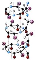
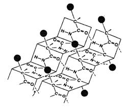
The primary structure of tens of thousands of different proteins is deciphered to date. For the definition of protein primary structure we find out amino-acid composition by hydrolysis methods. Then we determine the chemical nature of chain-terminal amino acid. The following stage is definition of sequence of amino acids in a polypeptide chain. For this purpose we use selective partial (chemical and enzymatic) hydrolysis. Also X-ray crystallographic analysis can be used. We can use the data on DNA sequence, because information about the synthesis of protein recorded in DNA. Secondary protein structure is a configuration of a polypeptide chain, i.e. means of packaging of a polypeptide chain in certain conformation. This process proceeds not chaotically, but according to the program put in primary frame. One of the most common type of secondary structure is a -helix (fig. 1). Curling of a polypeptide chain happens clockwise. Stability of secondary structure is provided basically by the intramolecular hydrogen bonds which exist between backbone –C=O and H-N- groups. A certain contribution can be made by covalent bonds - peptide and disulfide. For each protein certain degree of helix is characteristic. Hemoglobin chains are 75% helical, pepsin – are only 30%. Type of the configuration of polypeptide chains found in proteins of hair, silk, muscles, is called b -pleated sheet. This structure is stabilized by intermolecular hydrogen bonds. Segments of a peptide chain settle down in one layer, forming a figure similar to sheet, accordion-like folded. Polypeptide chains can be parallel or antiparallel. b-pleated sheet can be formed also intramolecularly. Fig. 1. Secondary structure of protein: a) a -helix , b) b -pleated sheet. In nature there are proteins constitution of which does not correspond neither to β - nor a - structure, for example, collagen - the febrile protein of connective tissue in a human body and animals. Tertiary protein structure is packing of a polypeptide chain in certain volume. The first protein, which tertiary structure has been found out by X-ray crystallographic analysis is myoglobin of a cachalot (fig. 2). Tertiary structure of proteins is maintained by covalent bonds (disulfide bonds). But the dominant role is played by noncovalent bonds (hydrogen bondes, electrostatic interactions of the charged groups (salt briges), intermolecular Van der Waals forces, hydrophobic interactions etc.). According to modern knowledge, tertiary protein structure after the end of its synthesis is formed spontaneously. The basic reason is interaction of amino acids radicals with water. Thus non-polar hydrophobic radicals of amino acids are dipped in protein molecule, and polar radicals are oriented towards water. The process of tertiary protein structure formation is called folding. In cells there are proteins called shaperons. They participate in folding. We know a series of hereditary human diseases the cause of which is infringement of folding process (pigmentosis, fibrosis, Mad Cow Disease). X-ray crystallographic analysis methods prove existence of levels of the structural organization of a protein molecule, which are mediate between the secondary and tertiary structures. The domain is a compact globular unit in a polypeptide chain (fig. 3). Many proteins are discovered (for example, immunoglobulins), consisting of different in structure and functions domains coded by different genes. Fig. 2. Myoglobin t ertiary structure Fig. 3. Globular domains in g- crystallin (protein of human eye crystalline lens ) All biological properties of proteins are bound to safety of their tertiary structure which is called native. The protein globule is not an absolutely rigid structure: reversible moves of parts of a peptide chain are possible. These changes do not break the general conformation of a molecule. Conformation of a molecule of protein is influenced by ionic strength of solution, interaction with other substances, рН. Any influences leading to infringement of native conformation of a molecule, is accompanied by particulate or full loss by protein of its biological properties. Quaternary protein structure. Formation of quaternary protein structure is packing in space of separate polypeptide chains into an organized whole. These chains may have identical or different primary, secondary or tertiary structure. The protein molecule consisting of several polypeptide chains, is called oligomer, and each chain entering it - monomer. Oligomer proteins are more often constructed of an even number of monomers, for example, the hemoglobin molecule consists of two a- and two b-polypeptide chains (fig. 4). About 5% of proteins possess quaternary structure, including hemoglobin, immunoglobulins. The subunit constitution is common with many enzymes. a chain 1 b chain 1 b chain 2 a chain 2 Fig. 4. Hemoglobin molecule The protein molecules which are part of the protein quaternary structure, are formed on ribosomes separately and only after the end of synthesis do they form the general supramolecular structure. Protein gets biological activity only after the affiliation of its compositional monomers. Quaternary structure is stabilized by the same types of forces as tertiary, but no covalent bonds. Some researchers recognize existence of the fifth level of the structural organization of proteins. These are metabolons - multifunctional macromolecular complexes of different enzymes catalyzing the whole pathway of metamorphosis of substrate (synthetases of the highest fat acids, pyruvate dehydrogenase complex, and respiratory chain). |
Последнее изменение этой страницы: 2019-06-19; Просмотров: 258; Нарушение авторского права страницы